ActivitiesPublished in Surfaces and Interfaces: The role of alloying on texture evolution and solid-state dewetting of thin metallic films
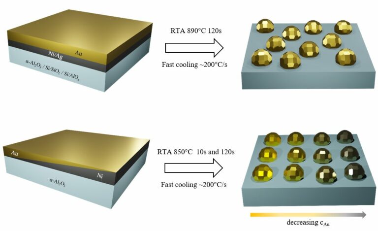
The paper “Influence of Au alloying on solid state dewetting kinetics and texture evolution of Ag and Ni thin films” by Martin Dierner, Johannes Will, Michael Landes, Christian Volland, Robert Branscheid, Tobias Zech, Tobias Unruh and Erdmann Spiecker has just been published in the Journal of Surfaces and Interfaces. Congratulations Martin and colleagues! Thin films […]The paper “Influence of Au alloying on solid state dewetting kinetics and texture evolution of Ag and Ni thin films” by Martin Dierner, Johannes Will, Michael Landes, Christian Volland, Robert Branscheid, Tobias Zech, Tobias Unruh and Erdmann Spiecker has just been published in the Journal of Surfaces and Interfaces. Congratulations Martin and colleagues! Thin films […]